mast cell tumor cat prognosis
Basal cell carcinomas are malignant tumors that occur most frequently in aged cats. These kinds of tumors are made up of many mast cells that release toxic granules creating allergic symptoms such as redness swelling or itching.
Mast Cell Tumors In Cats
The MTSS was not significantly different between the 4 groups.
. Surgical removal is the treatment of choice. Because tumors affect histamines this formation can result in side effects common in allergies like itching. For cats with Grade I mast cell tumors complete surgical excision carries an excellent prognosis with more than 90 percent of affected cats alive without disease after 4 years.
The prognosis for cats with visceral MCT varies depending on the location of the tumor. What Does a Mast Cell Tumor Look Like on a Dog. Feline mast cell tumors MCTs are frequently encountered in general practice.
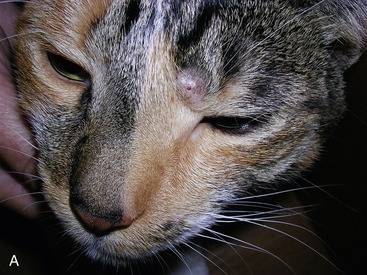
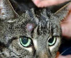
Typically they are shiny pink hairless nodules on the skin but there are many variations on this. Mast cell tumors MCTs are the most common malignant skin cancer in dogs and significant variability exists in their biological behavior. Unlike benign basal cell tumors these carcinomas are not usually raised up from the skin.
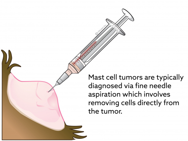
Grade III MCT are more aggressive tumors that are locally invasive and have a high rate of spread 50-90. Collapse Mast cell tumors on the skin cant be diagnosed just by looking at them because they. Grade II MCT are a more difficult group to.
None of the prognostic factors analyzed. Siamese cats appear to be predisposed to this form of MCT. Mast cell tumors MCTs are the most common skin tumor in dogs and the second most common skin tumor in cats.
The existing literature characterizes feline cutaneous mast cell tumors as mostly benign however approximately 20-25 of the cases show aggressive biological behavior with lymph node involvement and visceral metastasis. Respiratory distress can also be caused by pleural effusion or anemia which is present in up to 33 of cats. Two histologic grading systems are now described.
Because of this variable. They can be wide plaques or lumps. Mast cell tumors develop from specific cells of the immune system called mast cells which normally treat inflammation and allergic reactions in a dogs body.
Raised growth or bump Loss of hair in area of bump Redness or irritation of bump. Cats with visceral mast cell tumors present with signs of systemic disease such as. Well-differentiated and poorly differentiated.
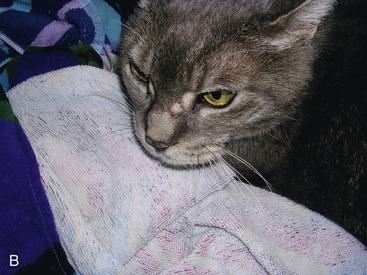
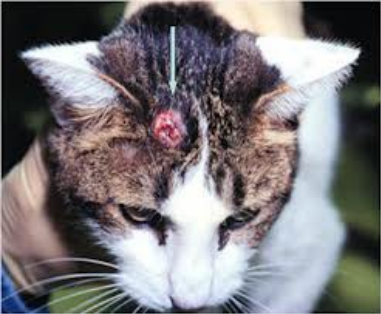
They often appear as ulcers on the head legs or neck. The symptoms of a cats mast cell tumor depend on whether the feline has cell tumors that are visceral or cutaneous. Cutaneous mast cell tumors present as lumps swellings or lesions in the skin or under the skin usually around the head and neck but sometimes elsewhere.
856 853 244 365 days for groups A B C and D respectively. For cats with Grade II mast cell tumors there is a wide spectrum of biological behavior but approximately 50 percent of affected cats survive for 4 years without tumor recurrence. In dogs MCTs can have either a benign or malignant behaviour and this is largely dependent on histologic grade.
A positive prognosis The prognosis for most cats with MCTs of the skin is very good. Symptoms of Mast Cell Tumor in Cats Like many tumors mast cell tumors will begin with raised bumps or growths with an escalation of symptoms depending on the severity of the condition and whether the tumor is cancerous. Mast cell tumors appear on cats as small firm hairless raised bumps or lumps on the skin.
Your cat may or may not have some itching in those areas depending on how it reacts to histamine. Treatment and prognosis can vary dramatically with location and histologic classification. Mast cell tumors typically affect older cats.
Grade I MCT act in a benign manner and most can be cured with surgical removal. Mast cell tumors were historically classified as one of three different histologic grades grades I II and III. If a mast cell tumor is affecting internal organs or the chemicals in the cells are being released in the body you might see these symptoms.
Lethargy and loss of energy. Pathologists divide mast cell tumors into two forms. Based on how the cells appear under the microscope well-differentiated what a more normal mast cell looks like or poorly-differentiated what a very abnormal mast cell looks like the disease progression and prognosis may vary.
This is why its important to regularly check your dog for lumps or growths and discuss any. Mast degranulation is usually episodic with systemic mastocytosis and clinical signs include GI ulceration uncontrollable hemorrhage altered smooth muscle tone hypotensive shock and respiratory distress. As mentioned above a standard indicator of cutaneous mast cell tumors is hard hairless bumps on the skin.
The well-differentiated tumors tend to behave. There is no grading system available in cats to distinguish these malignant cutaneous mast cell tumors from benign forms. The cause of these tumors is currently unknown and the tumors can develop anywhere on your dogs body.
Vomiting Depression Anorexy Weight loss Diarrhea Hyporexia Respiratory distress if pleural effusion Splenomegaly enlarged spleen Ascites Hepatomegaly enlarged liver Anemia 14-70 Mastocytosis 31-100. Most MCTs are cured with appropriate local therapy but a subset shows malignant behavior with the potential to spread to lymph nodes liver spleen and other areas and to thus become a systemic cancer. One study found the average age was 10 years.
Loss of appetite and weight loss. They spread forming new ulcers. Median tumor specific survival MTSS was.
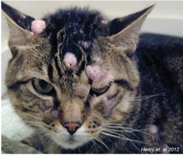
Surgical removal of the tumor usually results in a cure with little chance of recurrence at the surgical site or of spread to other sites in the body says Dr. Up to 14 dogs and 50 cats have multiple MCTs. The clinical signs of feline mast cell tumors depend on the location of the tumor.
The older three grade. MCTs are the most common splenic tumor second most common skin tumor and third most common intestinal tumor in cats. Persians are more prone to them.
However comparing cats that had splenectomy A and B versus those that did not C and D the MTSS was 856 and 342 days respectively p0008.

Mast Cell Tumors Animal Medical New City
Mast Cell Tumors Veterian Key
Mast Cell Tumors In Cats Veterinarian In Las Vegas Nv

Eyelid Mass With Conjunctival Periocular Swelling In A Cat Clinician S Brief
Mast Cell Tumors In Cats
Mast Cell Tumors Veterian Key

Periocular Cutaneous Mast Cell Tumors In Cats Evaluation Of Surgical Excision 33 Cases Semantic Scholar

Animal Surgical Center Of Michigan Veterinarian In Flint Mi

Mast Cell Tumors In Cats
Mast Cell Tumors In Cats Vca Animal Hospital

Pdf Diagnosis And Treatment Of A Feline Oral Mast Cell Tumor Semantic Scholar

Table 1 From Mast Cell Tumors In Cats Semantic Scholar

Animal Surgical Center Of Michigan Veterinarian In Flint Mi

Mast Cell Tumor Mastocytoma In Cats Petmd

10 Common Skin Tumors In Dogs And Cats Petplace
Mast Cell Tumors Veterinarian In Montgomery Al Animal Hospital Of Montgomery
Feline Cutaneous Mast Cell Tumors

Mast Cell Tumors In Cats Symptoms Diagnosis Treatment All About Cats
Mast Cell Tumors Veterinarian In Montgomery Al Animal Hospital Of Montgomery